An emergency medicine education platform for clinicians and trainees — for educational use only, not a substitute for clinical judgement or formal medical advice.
EDAITUTOR
ED Reference · Paeds Rashv0.1 · illustratedEducational use only
The Child with a Rash
A morphology-first read for the ED — does it blanch? · purpura vs petechiae · the febrile rash — and the can't-miss diagnoses hiding behind a fever.
Glass testPurpuraMeningococcal
🔴 Rash + unwell = dangerous until proven otherwise
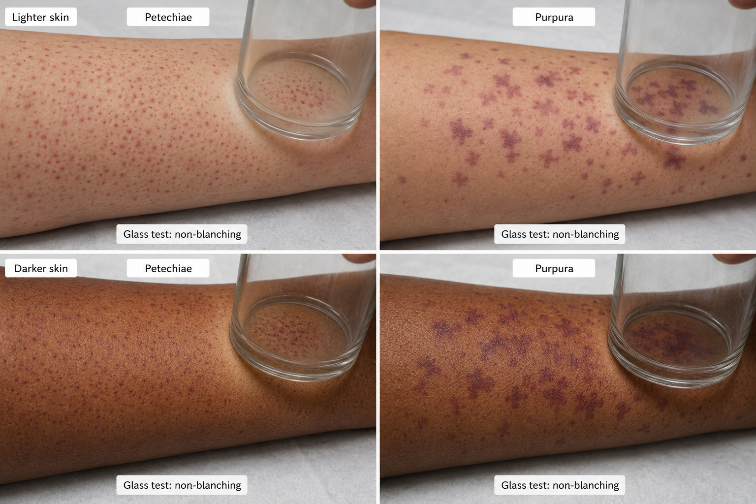
The one question that changes everything: does it blanch? A non-blanching rash in a febrile or unwell child means you exclude meningococcal disease & sepsis first. Purpura (>2 mm, confluent) is the real red flag — not scattered petechiae. Fully undress the child, do the glass test, and get senior review for any petechiae or purpura. If the child is unwell — bloods, cultures, meningococcal PCR and IV antibiotics, now.
Morphology
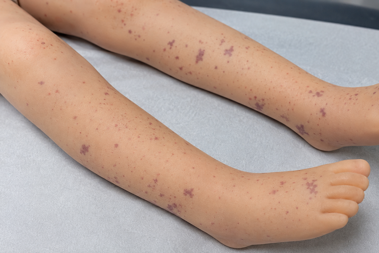
Petechial / purpuric — non-blanching, the high-stakes bucket
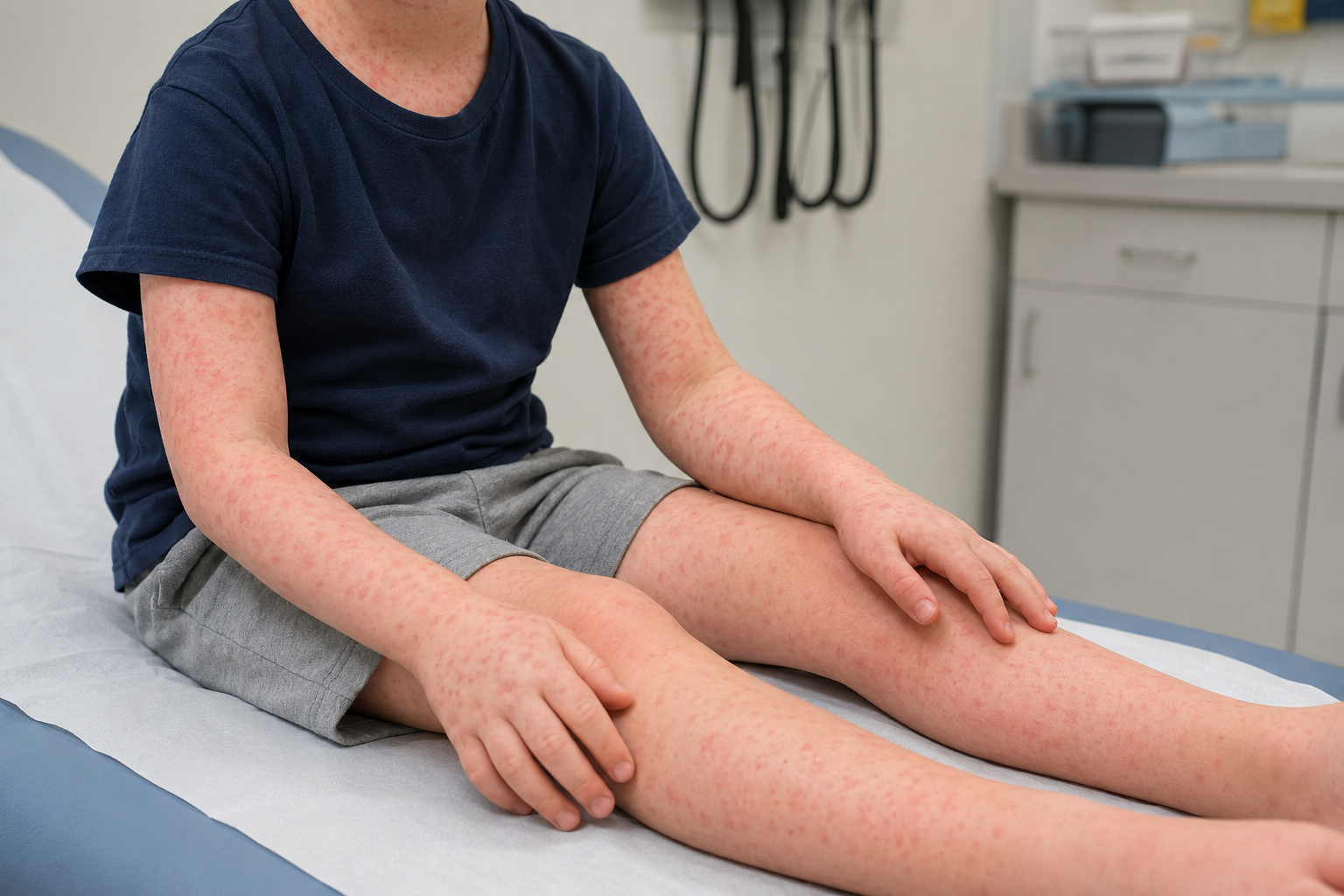
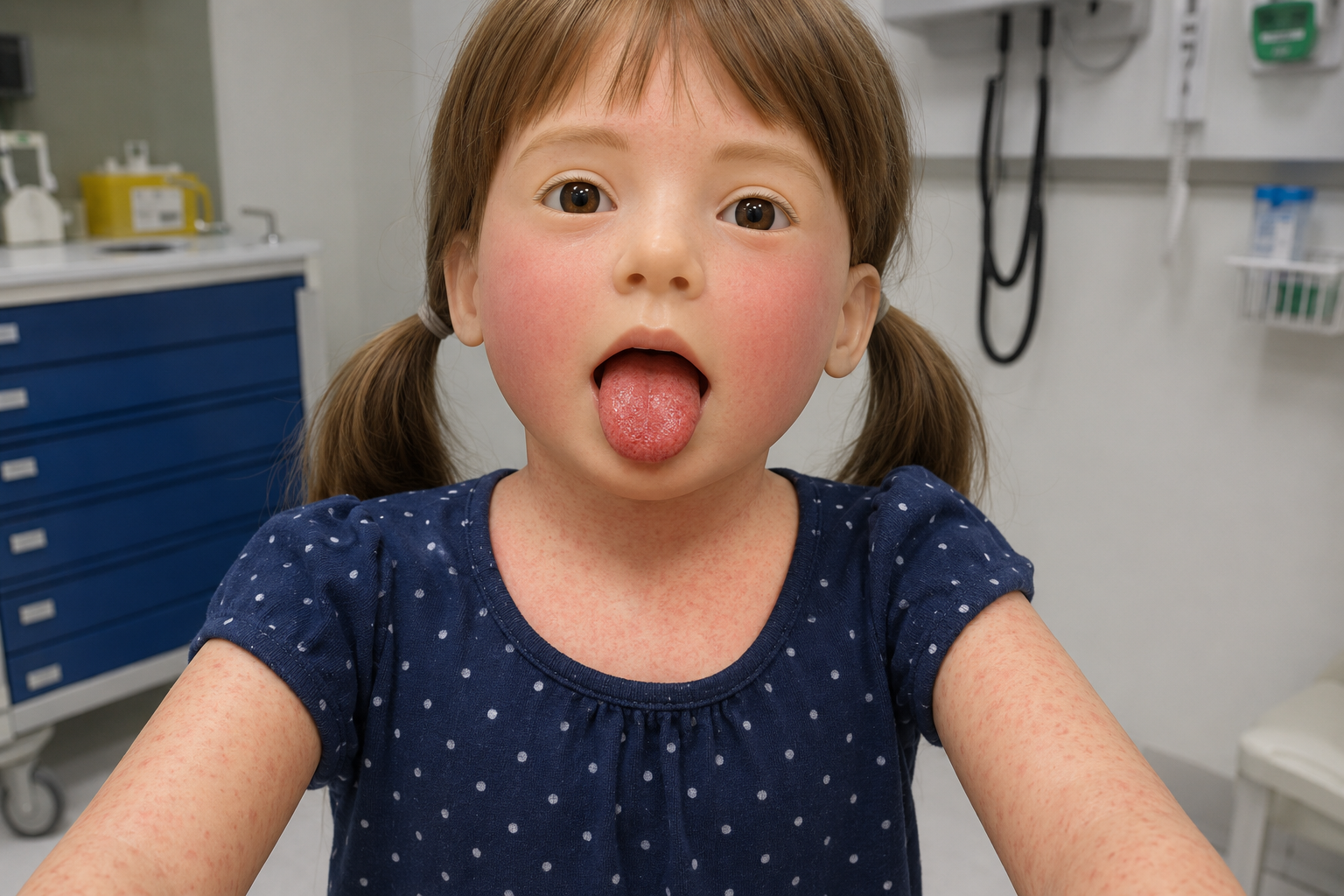
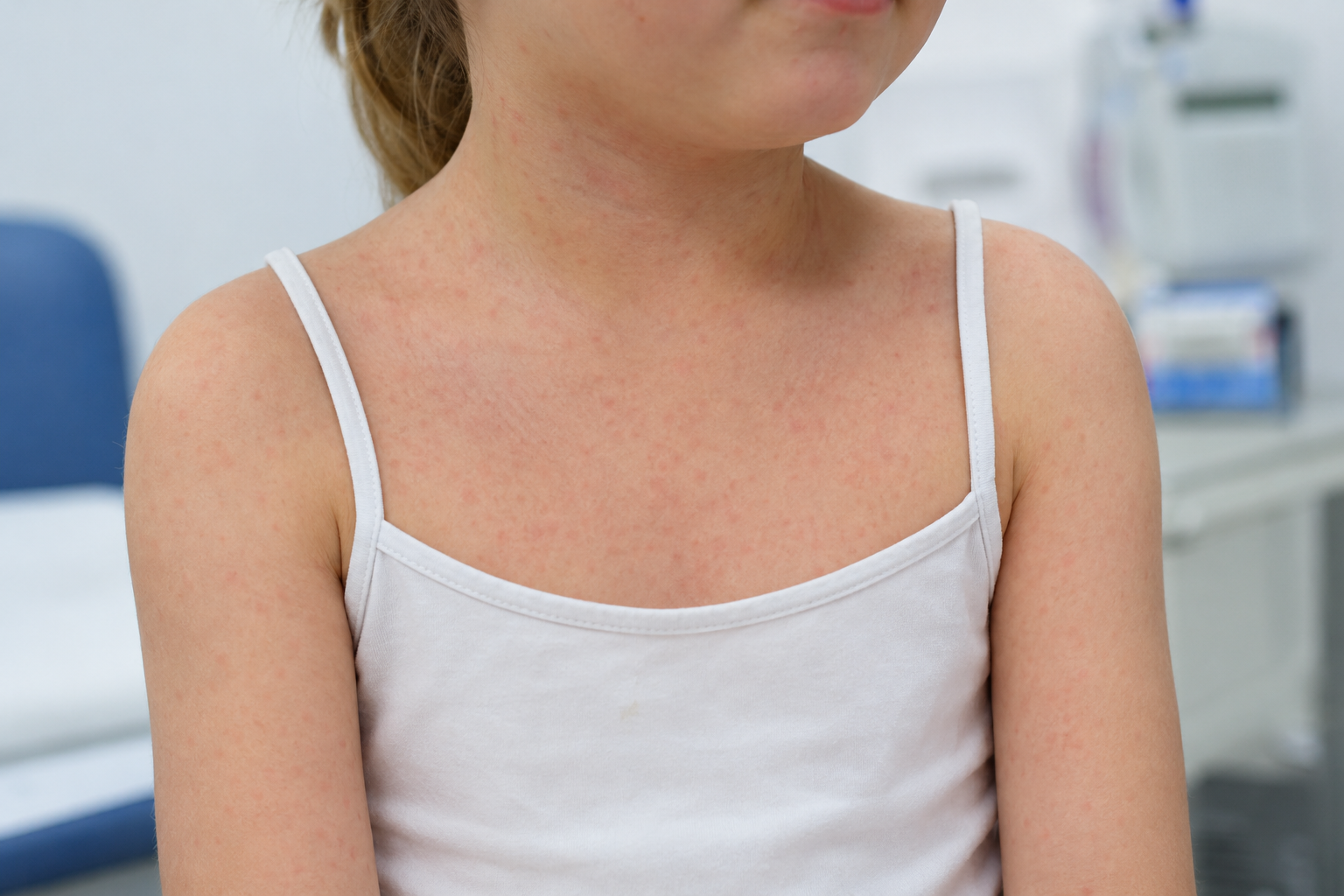
Maculopapular — flat + raised, blanching, the viral crowd
IV access — IM or IO if difficult; don't delay the dose
🟢 doseCeftriaxone 100 mg/kg (max 4 g) IV. Give as soon as invasive infection is suspected.
🟢 RCH · CHQ
The four morphologies — tap any image to enlarge
1Petechial / purpuric
Pinpoint to confluent, non-blanching lesions. The high-stakes bucket — think meningococcal, HSP, ITP.
Tap to enlarge
Clinical image · educational reference
2Maculopapular
Flat macules + raised papules, blanching. The viral crowd — and the classic drug-versus-viral trap.
Tap to enlarge
Clinical image · educational reference
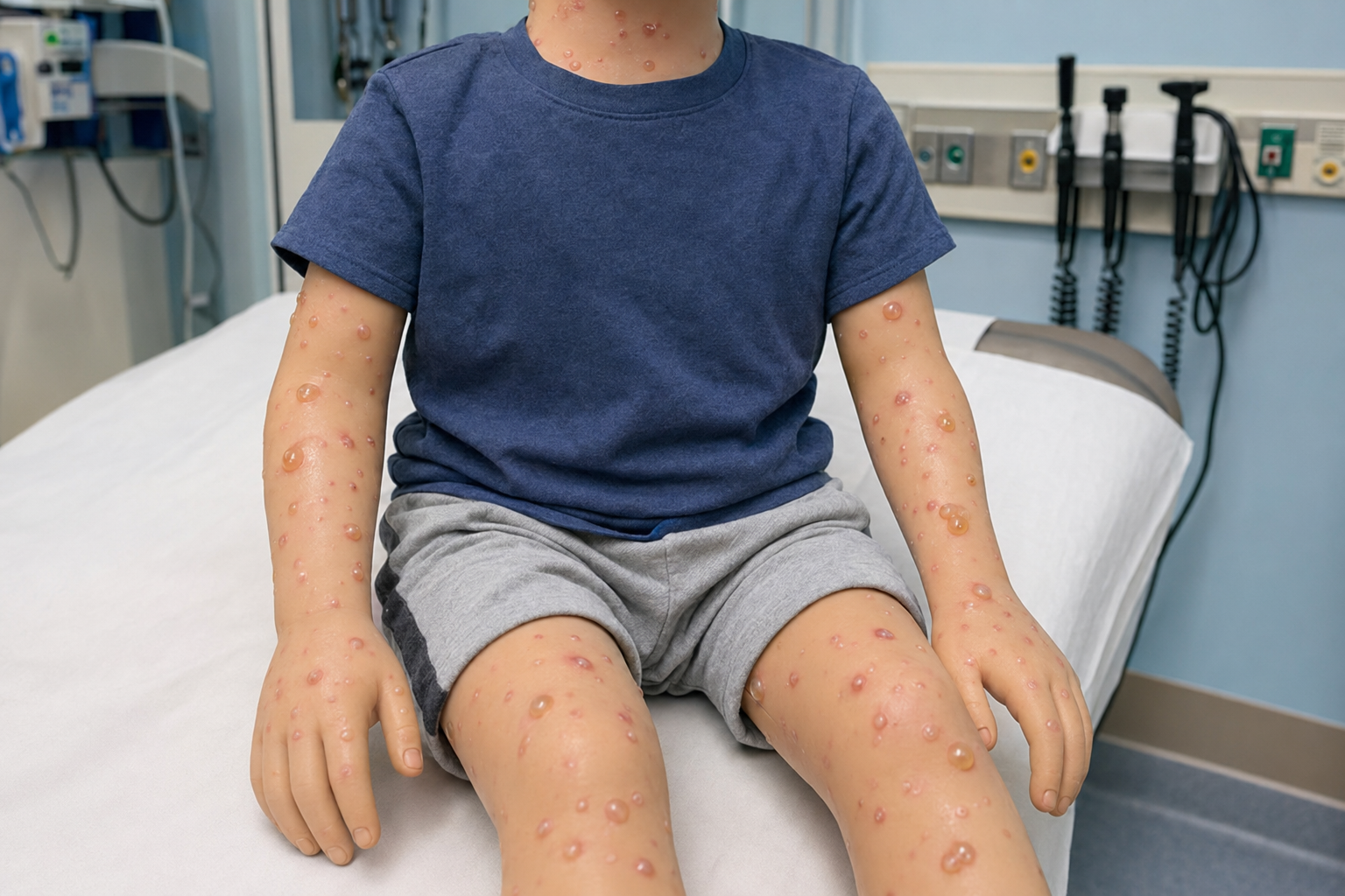
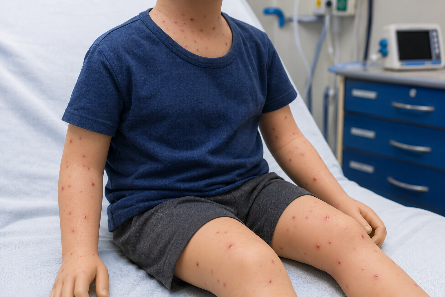
3Vesiculobullous
Fluid-filled vesicles or bullae — chickenpox and HFMD, but also SJS/TEN when skin is painful.
Tap to enlarge
Clinical image · educational reference
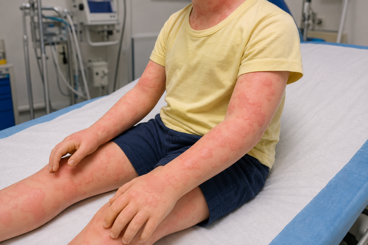
4Erythematous / urticarial
Migrating, blanching wheals that itch — the single most common rash reason for an ED visit.
Tap to enlarge
Clinical image · educational reference
⚪ sort, then nameGet the morphology and the child's appearance first; the named diagnosis follows. Images are external educational references, not produced by ED AI Tutor.
The non-blanching lane — exclude the life-threats first
◆Petechiae vs purpura + the tumbler test
Purpura (larger, confluent, >2 mm) carries far more risk than scattered petechiae — and the glass/tumbler test tells them apart from a blanching viral rash.
Tap to enlarge
Clinical image · educational reference
Meningococcal disease
Don't miss
Fever + petechiae/purpura, poor perfusion, altered conscious state. The rash is absent early in up to half of cases — its absence never reassures.
Treat first, investigate later
Henoch–Schönlein purpura
IgA vasculitis
Palpable purpura, lower-limb / buttock predominant, in a well child + abdominal pain, arthritis or renal signs.
Check BP + urinalysis
ITP & haematological
Well, afebrile
Widespread petechiae, bruising, mucosal bleeding without fever suggests thrombocytopenia.
FBC + film before reassuring
Non-accidental injury
Always exclude
Unexplained bruising or petechiae in a non-mobile infant, or a history that doesn't fit.
Safeguard + senior review
Mechanical petechiae
Benign mimic
Petechiae above the nipple line after coughing, vomiting or crying, in a well, afebrile child.
Reassuring distribution
The PiC study
What changed
Post men-B/C vaccine, invasive disease in fever + non-blanching rash fell from ~10–20% to ~1%. Meningococcal PCR stays positive for days after antibiotics.
Purpura > petechiae
⚪ modern messageIn a well, vaccinated child, isolated petechiae is no longer an automatic red flag — but purpura, an unwell child, or an evolving rash always is.
Febrile rashes that blanch — but still bite
Kawasaki disease
Time-critical
Fever ≥5 days + ≥4 of: conjunctivitis, lip/oral changes, rash, extremity changes, cervical node >15 mm. Coronary risk if missed.
SJS / TEN
Derm emergency
Target lesions, dusky/blistering centres, mucosal involvement and skin pain. Usually drug-induced — stop the drug, escalate.
Toxic shock syndrome
Toxin-mediated
Diffuse "sunburn" erythroderma + fever + shock, later desquamation. Hunt the source.
Rickettsial / spotted fever
Exposure history
Fever, headache, rash to wrists/ankles/palms/soles that may turn petechial. Ask about ticks and travel.
Necrotising fasciitis
Pain out of proportion
Erythema with pain out of proportion, rapid spread, toxicity. Skin signs lag — trust the pain.
Pain out of proportionErythema with pain beyond the visible findings → necrotising fasciitis.
Mucosal + target/bullaeEye, mouth or genital involvement with blistering → SJS/TEN.
Rapidly evolvingA rash spreading or changing in front of you.
Ill child, minor rashSystemic illness that outweighs the rash — trust the child, not the spots.
Newborn petechiaeAny petechiae in a neonate → prompt senior review.
🟢 RCH · DFTB · CHQ
ED AI Tutor — ED Reference Series · v0.1 · For clinician education only. Not a substitute for clinical judgement, local guidelines or senior oversight. Any child with a rash must be assessed in the context of the full clinical picture.
Paediatric rash · clinical image
Tap the image to zoom · tap again to fit. External educational reference. ⌨ Esc to close.